Contents: [Two Tilts]
[Fringe Visibility]
[Other Stuff] 

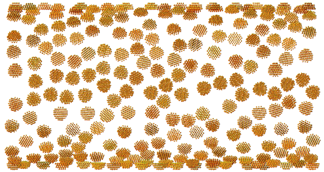
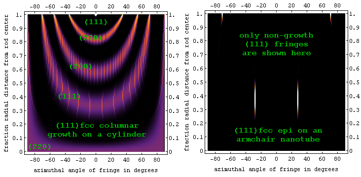
An application of lattice fringe visibility theory to the study of ordered particle growth on nanocylinders is described in this paper on "Lattice fringe signatures of epitaxy on nanotubes" to cond-mat/0603312.


These model predictions are easy (if tedious) to test against angle/position measurements of fringes from individual particles. Digital darkfield techniques provide an alternate way to compare model and experiment.
Sound Bytes: tilt to hide | nano-lattice
collimation | now you see it, now you don't Atom scale tunnels through a crystal become easier and easier to see as the crystal becomes thinner and thinner. This paper examines these effects for crystals in the 2 to 30 nm size range, both experimentally and theoretically working toward rules which allow one to predict quantitatively how crystal size and orientation alter the lattice fringes seen in an electron microscope. The result: Ball-shaped fringe visibility maps that allow one to answer a whole bunch of quantitative questions./pf 2005jan31
Headings: REL spots | lattice fringe
visibility | HRTEM | nanocrystal goniometry
Copyright (2005) American Institute of Physics (AIP), this article may be downloaded for personal use only. Any other use requires prior permission of the author and the AIP. It appeared in Journal of Applied Physics 98 (2005) 114308, the Dec 19, 2005 issue of Virtual Journal of Nanoscale Science and Technology, and is more generally available at this URL.
In context of this paper, here are a bunch of online-interactive fringe visibility and Kikuchi maps. Let us know if you find errors, or have other structures you'd like to see mapped.
Here are arXiv PDFs for versions [2] and [1].
Here is updated draft version [2+] , and version [1], both with higher resolution figures.
BiBTeX reference:
@article{wqpf.ncso,
author = "P. Fraundorf, Wentao Qin, P. Moeck, and Eric Mandell",
title = "Making sense of nanocrystal lattice images",
journal = "Journal of Applied Physics",
volume = "98",
year = "2005",
pages = "114308-1--114308-10",
eprint = "arXiv:cond-mat/0212281"}


This paper considers how nano-humans might determine the 3D structure of crystals, if they were able to re-orient the crystal in their hand while trying to peer down along tunnels between the columns of atoms which run in various directions. In that case, they could formulate rules (protocols) for everyone to use in recognizing a face-centered cubic crystal, a body-centered cubic crystal, etc. Precisely this type of manipulation (imagine a hand which can only tilt the crystal through a limited angle, over one or two fixed axes) is becoming increasingly possible with modern day atomic resolution transmission electron microscopes. It is therefore time to summarize and refine strategies, including those discussed here./pf 2002oct02
Sound Bytes:  hand-analysis for nano-geologists | 3D lattices from
images at two tilts | touring crystals on $5/eV
Elsevier's published version is available through ScienceDirect, but posted with permission here only in the Book section of our limited-access Nanoscale Science and Technology Blackboard.
wqpf.lptt PDF draft
BiBTeX reference:
@article{wqpf.lptt,
author = "Wentao Qin and P. Fraundorf",
title = "Lattice parameters from direct-space images at two tilts",
journal = "Ultramicroscopy",
volume = "94",
number = "3-4",
pages = "245--262",
year = "2003",
eprint = "arXiv:cond-mat/0001139"}

A subset, and often the whole, reciprocal lattice of a crystal can be inferred from the xyz coordinates of three non-coplanar reciprocal lattice vectors. Such coordinates may be obtained from electron-phase or Z contrast images taken at two tilts, provided that one image shows two non-colinear lattice periodicities, and the other shows a periodicity not coplanar with those two. We show here how to find, and implement, protocols for measuring the 3D parameters of any lattice type in this way. Particularly for cubic crystals with cell side greater than twice the appropriate resolution limit, we show that orthogonal +/-15 and +/-10 degree tilt ranges may allow one to measure 3D parameters of all varieties in a specimen, from only two well-chosen images. The strategy is illustrated by measuring the lattice parameters of a 10nm WC_{1-x} crystal in a plasma-enhanced chemical-vapor-deposited thin film.

Title: On-line determination of nanocrystal lattice parameters
Authors: W. Qin1, 2 and P. Fraundorf1
1Physics/Astronomy Department and Center for Molecular
Electronics, University of Missouri-St. Louis, St. Louis, MO 63121
2Process and Materials Characterization Lab, Digital DNATM Labs, Motorola Inc.,
MD EL622, 2100 E Elliot Road, Tempe, AZ 85284
Selected slides from the summer 2003 presentation
Abstract: The three-dimensional lattice parameters of a selected crystal can be inferred from lattice image information on three sets of non-parallel lattice planes. Today, with sufficiently wide tilt-capability, such data can come from phase-contrast (or less easily Z-contrast) images taken along two low-index zone axes of the crystal (cf. Ultramicroscopy 94, p. 245-262). Higher spatial resolution in images will lessen the requirement for wide-angle tilting, but also increase the geometric complexity of the task due to the involvement of lower symmetry orientations. In addition to some "fancy inverse crystallography" for the design of tilt protocols, our experience so far also suggests that issues of tilt-stage precision, on-line computer support, and off-axis fringe visibility as a function of specimen thickness will have to be considered before routine on-line determination of the lattice parameters of an arbitrary nanocrystal becomes possible in practice.recent submissions to Microscopy and Microanalysis 2004.
notes by Peter Möck at Portland State on TEM goniometry.
earlier TEAM (aberration-corrected microscopy) presentations.
Papers on fringe visibility maps and lattice parameter uncertainties in PDF form, submitted to MSA 2001.
Wentao's thesis in PDF form.Papers on tilt (p. 1040-1041) and probability (p. 1038-1039) in PDF form, for MSA 2000.
An early version of a paper on direct-lattice crystallography, archived at Los Alamos in e-print form.
Our first paper on stereo analysis of 2D electron diffraction data.

Above: a fringe-visibility map for 2nm diameter elemental fcc (e.g. Al) crystals (lattice parameter near 0.4nm) in a scope capable of 0.14nm resolution.
Below: Fringe visibility maps for some of the larger fringes visible from body-centered, face-centered, and diamond face-centered lattices. Note the dominance of crossed (110) fringes at the three-fold <111> zone in the body-centered case, the dominant crossed (111) fringes at the two-fold <110> zone in the face-centered cases, and the wider disparity between largest and next-to-largest spacings when the diamond glide is added to the lattice.


A paper in PDF form for the Microscope Society of America Meeting in Summer 1999.
A number of opportunities for wider application, as well as adaptation for on-line use in computer-based microscopes, present themselves downstream...